 Collaborators: Christopher Rodesch, PhD, Mike Redd, PhD
Departments: Neurobiology and Anatomy, Fluorescence Microscopy Core
Project
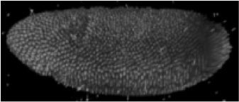
The HSC Fluorescence Microscopy Core is in the process of assembling a Single Plane Illumination Microscope (SPIM) for the real time visualization of large format specimens such as zebrafish and developing mouse embryo explants. The hardware design prototype allows for production of 3D data sets spanning an entire zebrafish embryo throughout development in a way that dramatically reduces photodamage and increases axial resolution. SPIM is deemed a valuable technology to pursue because it will provide a level of 3D resolution not available using confocal microscopy or other techniques. Traditional illumination schemes have a 2x reduction in resolution parallel with the axis of illumination when compared to the lateral resolution perpendicular to the illumination. DiSPIM images a sample from two of more angles and uses the separate lateral information from these data sets to define an isotropic rather than asymmetric resolved image volume. This is particularly important for relatively thick samples such as zebrafish or mouse embryos where the samples are as thick in the axial plane as they are broad in the lateral view.
BIDAC Contact: Clement Vachet
BIDAC Expertise
Assemble a Single Plane Illumination Microscope (SPIM) for the real time visualization of large format specimens such as zebrafish. Provide software tools that are more user friendly and more advanced than those currently available.
Progress
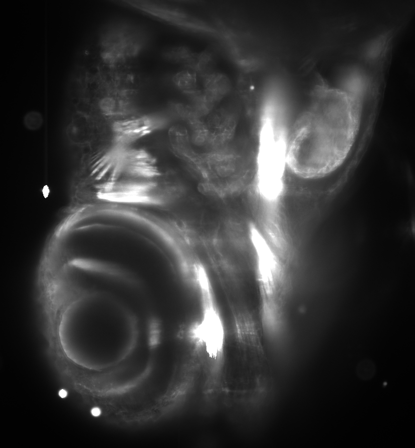
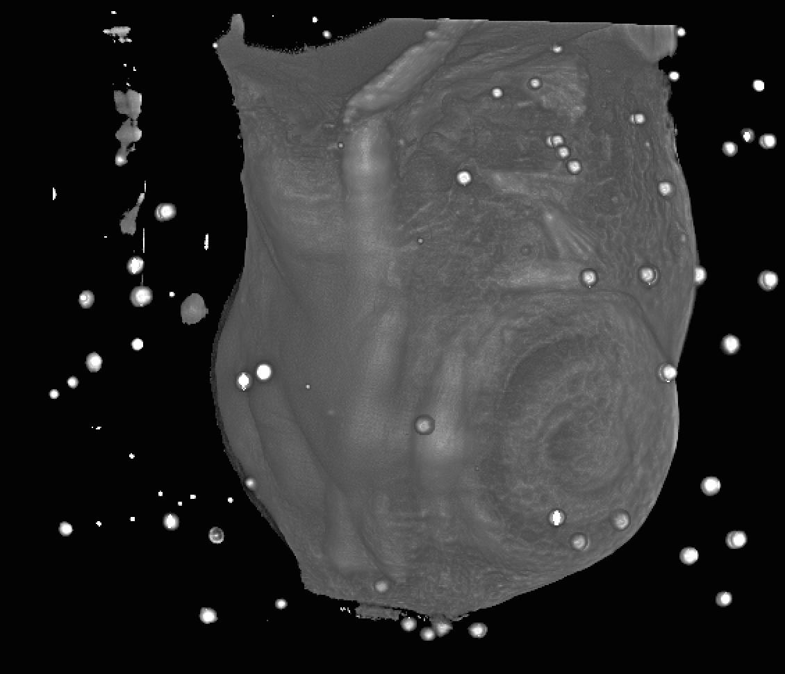
We provided advice on image acquisition strategies for data analysis needs, and enabled 3D visualizations of acquisitions via SCI software solution FluoRender. We have been investigating 3D reconstruction of multi-angle data acquisition via an open-source image processing framework (in progress).
General contribution
Open-hardware open-software SPIM solution delivers high signal-to-noise isotropic 3D images of large specimen from different angles in an extended time-lapse, which is currently hard to achieve with any other microscopy technology.
Presentations
OpenSPIM report (.pdf)
 |
 |
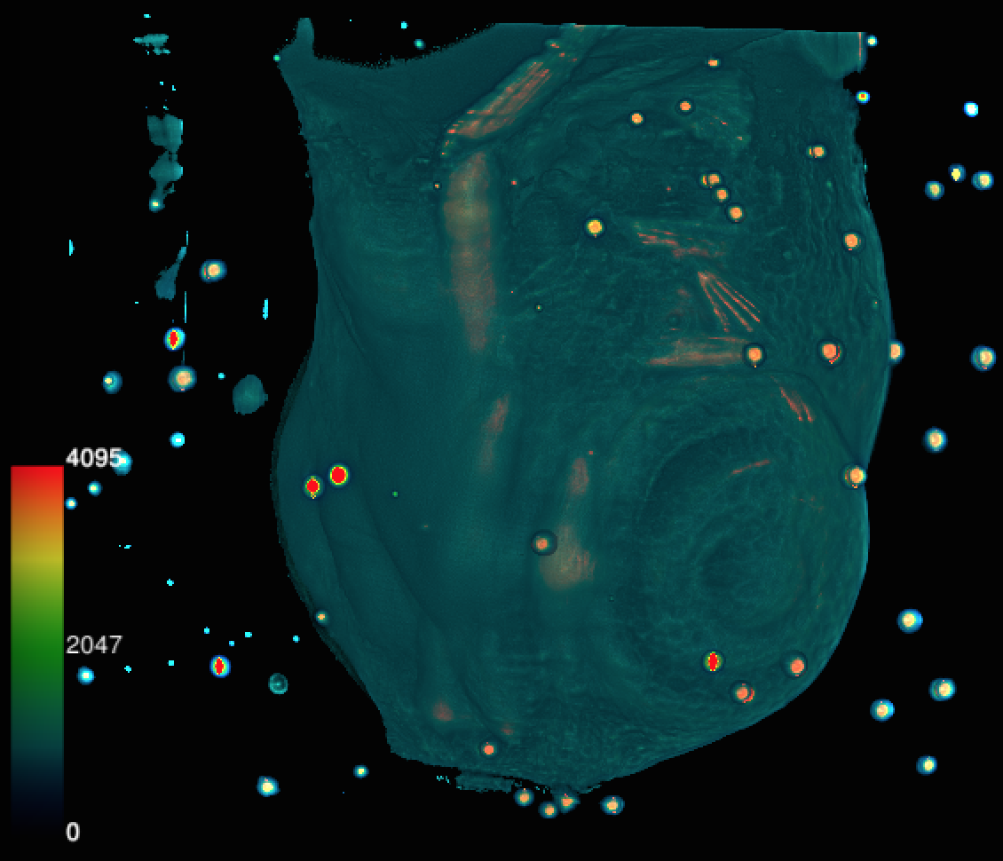 |
| ImageJ - 2D view | FluoRender - 3D view | FluoRender - colored 3D view |